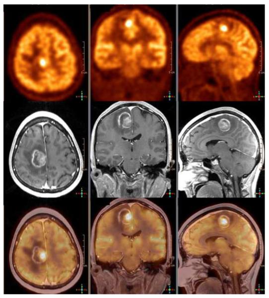
Figure 7.
49 year old man with left leg weakness. Axial, coronal, and sagittal 18F-FDG PET (top row), contrast enhanced 3D SPGR (middle row) and co-registered PET/ MR images demonstrate an asymmetrical enhancing medial precentral gyrus lesion with a cystic component and prominent surrounding white matter edema. Hybrid images document significantly increased glucose metabolism within the most enhancing region, a Grade 4 glioma.

