
Fig. 4.
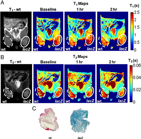
In vivo lacZ gene reporter activity of C3-GD. MRI of a representative nude mouse with wild type MCF7 tumor (left, dotted) and lacZ transfected MCF7 tumor (right, solid). A) Baseline T1 weighted image and T1 maps obtained before, 1 h after, and 2 h after direct intra tumoral injection of 15 mM C3-GD and 5 mM FAC (top row) and B) corresponding T2 weighted images and T2 maps (bottom row) showed decrease in the relaxation times in lacZ transfected tumors. C) X-gal and Nuclear fast staining of slices (whole mount) from the same wild type MCF7 (left) and MCF7-lacZ (right) tumors showed β-gal activity (intense blue stain from X-gal) for the MCF7-lacZ tumor section only.