
Figure 5. Laterality defects in dendritic spine morphology of the β2m-deficient hippocampus.
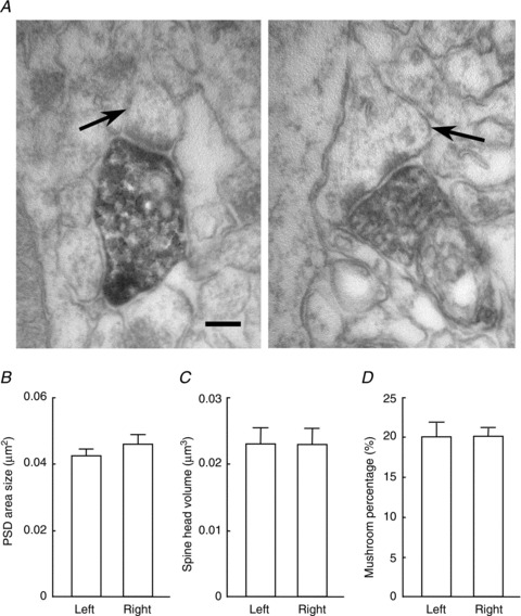
A, electron micrograph of CA1 pyramidal cell synapses in the middle third of the stratum radiatum in the β2m-deficient hippocampus. GFP-expressing lentivirus was injected unilaterally into the CA3 pyramidal cell layer. Axons and the terminals of ipsilateral CA3 neurones were heavily labelled for GFP. Arrows indicate thin (left) and mushroom-type (right) spines making contact with GFP-labelled axon terminals. Scale bar: 300 nm. B–D, average postsynaptic density (PSD) area (B), spine head volume (C) and percentage of mushroom-type spine (D) were compared between the left and right CA1 pyramidal cell synapses of the β2m-deficient hippocampus. No significant differences (P > 0.05) in laterality were observed in the three ultrastructural parameters. Error bars represent SEM.