
Figure 2. Immunocytochemistry.
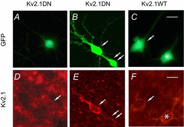
We stained organotypic slices with the Neuromab Kv2.1 monoclonal antibody (K89/34). Slices contained non-transfected cells and cells with GFP fluorescence, indicating biolistic transfection with GFP plus the Kv2.1 DN construct (A, D and B, E), or GFP plus overexpression of Kv2.1 WT (C, F). Note the differing exposures in D vs. E and F. In E and F we focused on the enhanced, brighter staining of the transfected cells at the expense of viewing non-transfected cells, and thus the background is dimmer. Non-transfected cells (no GFP fluorescence) showed a typical patchy distribution of clusters of channels on the soma and proximal apical dendrites (D). The staining pattern was similar for some cells expressing the Kv2.1 DN (e.g. cell with arrow in D, cell marked with double arrow in E). Other cells transfected with the Kv2.1 DN (cell marked with single arrow in E) and most cells transfected with the Kv2.1 WT appeared more intensely stained with a more continuous pattern. The Kv2.1 WT cells in F (arrow, asterisk) showed intense staining compared to non-transfected cells but retained a clustered distribution. Scale bars = 25 μm.