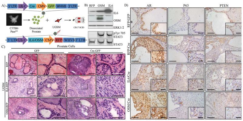
Figure 1. Paracrine expression of IL6 or OSM synergizes with epithelial loss of PTEN to promote invasive adenocarcinoma.
A) Diagram of regeneration and transformation process with lentiviral vector diagrams.
B1–3) Western analysis of UGSM cells infected with control, IL6 and OSM vectors showing heightened expression of IL6 and OSM in their respective cell lines, with a mild increase in IL6 expression in OSM-infected UGSM. Loading control is ERK1/2.
B4–5) Secretion and activity of the IL6 and OSM cytokines was confirmed by treating serum-starved 3T3 cells with conditioned media from infected UGSM. Increased phosphorylation of STAT3 in cells treated with IL6 and OSM conditioned media indicates functional activity. Imaged using Licor Odyssey CLx with Image Studio software.
C) Representative histological sections of prostate regeneration and transformation following 6–8 weeks in vivo. Control grafts exhibit normal prostate epithelial architecture with PTEN-intact grafts expressing either IL6 or OSM exhibited reduced epithelial regeneration while IL6- and OSM-expressing grafts exhibited occasional mild, focal hyperplasia. Grafts with loss of PTEN alone exhibited PIN lesions formation with characteristic neoplastic growth into the luminal interior while tumor foci from PTENLOF with IL6 or OSM exhibited invasion into the surrounding mesenchyme. Scale Bars: 10X, 200 um; 40X, 100 um.
D) IHC analysis of AR and p63 in prostate regenerations. All prostate epithelial regenerations and tumor foci retained high expression of nuclear AR. Normal regenerations exhibited P63-expressing basal cells along the basement membrane and were detached from the membrane in PIN lesions present in PTENLOF grafts and PTENLOF grafts with IL6. High grade lesions present in PTENLOF with OSM exhibited loss of P63-expressing basal cells. Scale Bars: 20X, 100μm; 63X, 50 μm

