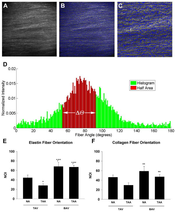
Figure 2.
Determination of matrix fiber orientation. A) Representative monochromatic image of collagen fiber matrix of a BAV-TAA patient using multi-photon microscopy. Magnification is 25X. B) Vector map (blue arrows) of fiber alignment for image in (A). C) Increased magnification of box indicated in (B) to illustrate fiber alignment vectors. D) Representative histogram of collagen fiber angle counts for BAV-TAA generated from multi-photon image in (A) to calculate the orientation index (Δθ). E–F) Normalized orientation indices (NOIs) for elastin and collagen respectively. Bars represent mean ± SEM, n=5 (TAV-NA), 8,9 (TAV-TAA), 6 (BAV-NA) and 10 (BAV-TAA) *Significant from TAV-NA, ** Significant from TAV-TAA, p<0.05.

