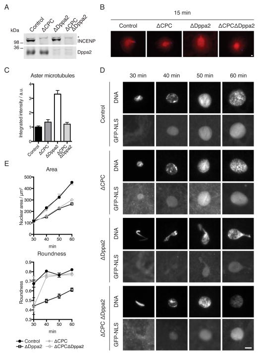
Figure 4. CPC depletion rescues ΔDppa2 phenotypes.
(A) CPC was depleted from extracts using anti-INCENP antibodies (Sampath et al., 2004) and depletion efficiency assessed by Western blotting. * indicates non-specific reactivity.
(B) Sperm nuclei were added together with calcium to metaphase extracts containing rhodamine-labeled tubulin (red). Sperm-associated asters were fixed after 15 min and stained with Hoechst 33342 (blue).
(C) Quantification of tubulin intensity from (B). Bars indicate mean and standard error of > 30 asters from each sample and are representative of 3 independent experiments.
(D) Sperm nuclei were added together with calcium to metaphase extracts supplemented with GST-GFP-NLS. Nuclei were fixed and visualized with Hoechst 33342.
(E) Quantification of nuclear area and roundness from (D). Bars indicate mean and standard error of > 30 nuclei from each sample and are representative of 3 independent experiments.
Scale bars, 10 μm.
See also Figure S4.

