Figure 3.
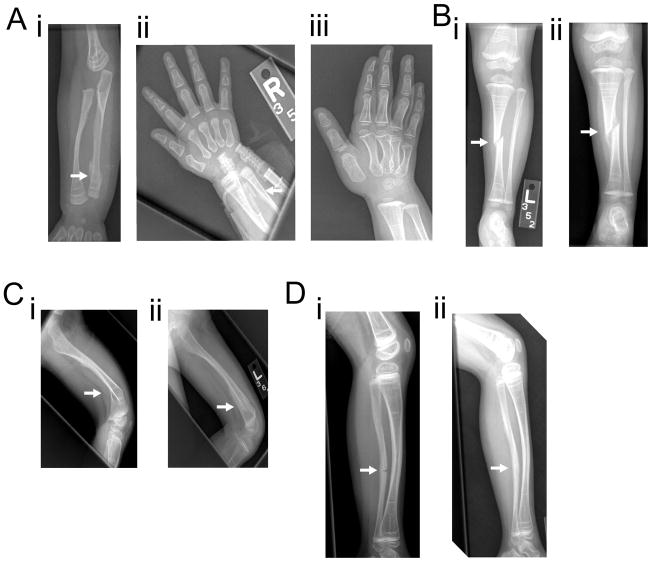
Figure 3A: i Right forearm radiograph at 8 months of age showing distal right ulnar fracture. Retained calcified cartilage at the metaphyses is noted, likely from bisphosphonate therapy. ii Right hand radiograph from 6 months later showing interval healing. iii Right hand radiograph 12 months after the occurrence of fracture shows healing at the fracture site.
Figure 3B: i Anteroposterior left leg radiograph at 2 years of age showing an acute oblique fracture of the mid-tibial diaphysis with one-half shaft-width lateral displacement. ii Repeat radiograph 1 month later shows an appropriate amount of callus has developed at the tibial fracture site.
Figure 3C: i Lateral left femoral radiograph at 2.5 years of age shows an acute fracture through the distal diaphysis of the left femur which is associated with approximately 40 degrees of posterior angulation. ii Follow up radiograph a month later shows interval callus formation and improved angulation.
Figure 3D: i Lateral left leg radiograph at 5 years of age demonstrates an acute non-displaced fracture involving the anterior cortex of the midshaft of the left fibula with associated bowing deformity. ii Repeat radiograph 6 months later shows healing at the fracture site.

