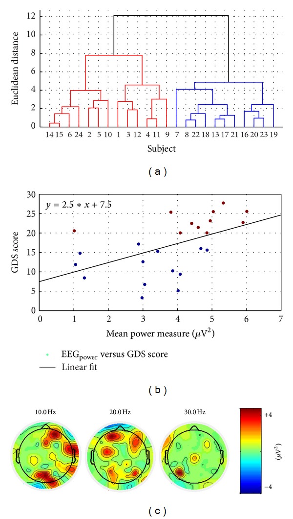
Figure 4.

(a) Dendrogram plot of the hierarchical cluster tree from the data set—mean power measure at O1, O2, and P8 electrodes averaged over 4–30 Hz versus GDS scores. (b) Scatter plot between GDS scores and mean power measures where marker colors blue for “nondepressed” and brown for “depressed” show the two hierarchical clusters based on the Euclidean distance. (c) Topographic maps of difference EEG spectra at 10 Hz, 20 Hz, and 30 Hz between “depressed” (GDS ≥ 15) and “nondepressed” (GDS < 15) QEEG groups.
