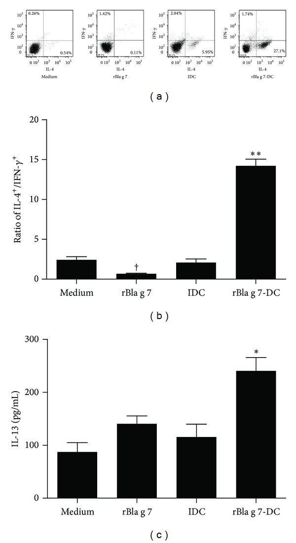
Figure 5.

Induction of Th2 polarization by rBla g 7-stimulated dendritic cell (DC)s. Isolated CD4+ T cells were cocultured with immature DCs (iDC), rBla g 7-activated DCs (rBla g 7-DCs), or rBla g 7-pulsed alone (rBla g 7) for 48 h at 37°C before being harvested for analysis. (a) Flow cytometry analysis of intracellular expression of IFN-γ and IL-4 by using the fluorescent-labeled antibodies. It was noticed that the number of IL-4+ cells in the lower right quarters of IDC and rBla g 7-DCs groups was increased. (b) The ratio of IL-4+ CD4+ T cells versus IFN-γ+ CD4+ T cells. (c) Levels of IL-13 in the culture supernatants determined by ELISA. The data were represented as the mean ± SEM for four separate experiments. *P < 0.05, **P < 0.01 in comparison with medium alone control. † P < 0.05 for decreased response in comparison with control.
