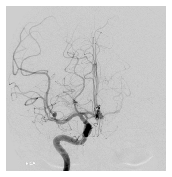
Figure 8.

Cerebral angiographic (DSA) image in AP view of right ICA at 6-month follow-up, demonstrating complete obliteration of the embolized aneurysm with retrograde filling of the normal artery distal to the aneurysm. The smaller aneurysm has shown significant reduction in size.
