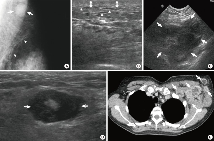
Figure 5.
A 43-year-old woman with diffuse large B-cell lymphoma. (A) Left mediolateral oblique mammogram shows axillary lymphadenopathy (arrows) and trabecular thickening (arrowheads). (B) Ultrasonography (US) scans of the left breast show skin thickening (arrows) and dilated dermal lymphatics (arrowheads). (C, D) US scans of the left axilla show enlarged lymph nodes (arrows). One node is indistinct and irregular in shape (C) and the other is circumscribed and ovoid (D). The nodes have cortical thickening. (E) Enhanced breast computed tomography image shows multiple-rim enhancing lymph nodes (arrows) in left axilla.

