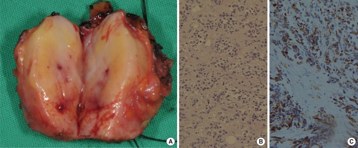
Figure 3.
Histologic findings of metastatic rhabdomyosarcoma in breast. (A) The rhabdomyosarcoma was oval and well-circumscribed, 2.2 cm sized in diameter. The cut surface showed focal hemorrhage with white central compartment encircled by yellow area. The small round cells are arranged in variably sized nest by fibrous tissue septa (H&E stain, ×200) (B) and strongly stained with desmin immunohistochemical stain (desmin stain, ×200) (C).

