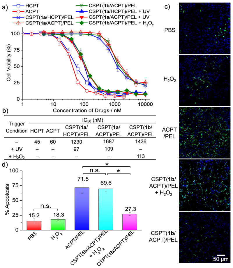
Figure 3.
(a, b) Cytotoxicity of CSPTs/PEL NPs in HeLa cells with or without trigger treatment was analyzed by microculture tetrazolium (MTT) assay. Triggering conditions: UV treatment (360 nm, 20 mW/cm2, 10 min) for CSPT(1a/HCPT)/PEL NPs and CSPT(1a/ACPT)/PEL NPs; H2O2 treatment (1 mM) for CSPT(1b/ACPT)/PEL NPs. The half maximal inhibitory concentration (IC50) values were determined by half-cell viability concentration from the MTT assay and summarized in the table. (c, d) BALB/c mice bearing subcutaneous 4T1 tumors received a single intratumoral injection of phosphate buffered saline (PBS), H2O2, ACPT or CSPT(1b/ACPT)/PEL NPs (0.5 mg ACPT equiv/tumor) with or without H2O2 (10 mM, 100 μL/tumor). H2O2 was administered intratumorally 1h after the injection of CSPT(1b/ACPT)/PEL NPs. The mice were sacrificed 48 h post injection. The 4T1 tumors were collected, sectioned and stained with deoxynucleotidyl transferase–mediated deoxyuridine triphosphate nick end (TUNEL) for apoptosis analysis. Representative images (c) and quantification by ImageJ (d) of TUNEL stains are shown. Scale bar: 50 μm. The apoptosis index was determined as the ratio of apoptotic cell number (TUNEL, green) to the total cell number (4′,6-diamidino-2-phenylindole (DAPI), blue) (20 tissue sections were counted per tumor; n = 4; data are represented as average ± SEM and analyzed by One-way ANOVA (Fisher) (*p < 0.05; n.s. = not significant)).

