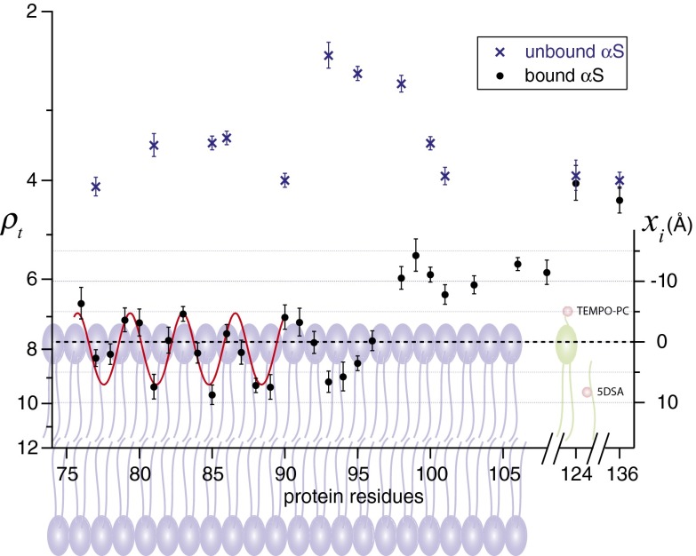
Fig. 3.
Retardation factor (ρt) at various residues of membrane-bound and free α-synuclein measured by ODNP. The distance (xi) of nitroxide radicals with respect to the phosphate group of the POPC/POPS bilayers is presented. Error bars represent SDs. The red curves are least-squares fits of data to a cosine function. The representative phospholipids and lipid spin probes are shown in the background. Nitroxide radicals of lipid spin probes are represented in red balls.

