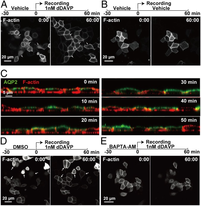
Fig. 6.
Vasopressin-induced calcium-dependent apical F-actin dynamics in mpkCCD cells. First (00 min:00 s) and last (60 min:00 s) images from recordings of apical F-actin in live and polarized mpkCCD cells in the presence (A) or absence (B) of dDAVP. (C) Time course of AQP2 and F-actin localization changes in mpkCCD cells following dDAVP treatment. First (00 min:00 s) and last (60 min:00 s) images from recordings of apical F-actin in mpkCCD cells pretreated with vehicle (DMSO) (D) or 50 μM BAPTA-AM intracellular calcium chelator (E) before the cells were exposed to dDAVP.

