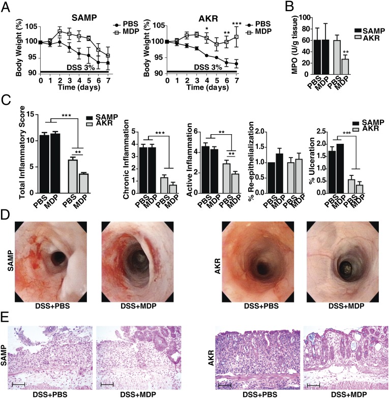
Fig. 1.
MDP administration in vivo reduces DSS colitis in AKR mice, but not in SAMP mice. SAMP and AKR mice were treated with 3% DSS in their drinking water for 7 d (n = 8–11 per group). At the early phase of colitis induction (days 0, 1, 2), mice were administered either MDP (100 μg, i.p.) or PBS daily. (A) Changes in body weight in SAMP and AKR mice administered MDP or PBS (two-way ANOVA repeated measures, MDP protective effect for AKR was significant at P = 0.023, but not for SAMP, P = 0.125). (B) Myeloperoxidase (MPO) activity calculated from the colons of treated mice (Kruskal–Wallis, P < 0.01, Dunn’s). (C) Colonic total inflammatory scores, as determined by the sum of chronic inflammation, active inflammation, percentage reepithelialization, and percentage of ulceration (one-way ANOVA, P < 0.001; pairwise Bonferroni). (D) High-resolution endoscopic images of the proximal colon after 7 d of DSS treatment show severe inflammation in both groups of SAMP mice (PBS and MDP) and mild inflammation (including slight vascular changes and mild granularity) in AKR control mice treated with MDP compared with PBS. (E) Representative histopathological sections show active, severe ulcers, adjacent regenerative crypts, active cryptitis, and increased inflammatory cells in the lamina propria of SAMP mice treated with PBS and MDP. Sections from AKR mice treated with MDP show regenerative colonic mucosa with focal mild, active cryptitis, and more minimal increased inflammatory cells compared with PBS-treated AKR mice. (Scale bars, 100 µm.) Data are represented as mean ± SEM. The single asterisk (*), double asterisk (**), and triple asterisk (***) denote significant differences at P < 0.05, P < 0.01, and P < 0.001, respectively. Results are representative of three independent experiments.

