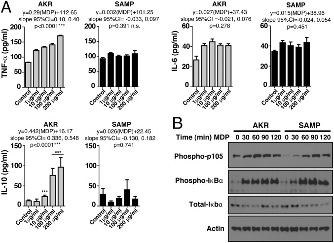
Fig. 3.
Impaired in vitro production of innate cytokines and NOD2 signaling in response to MDP in SAMP mice. (A) BMDMs isolated from preinflamed SAMP (4 wk old) and age-matched AKR control mice were incubated with different concentrations of MDP (1, 10, 100, 200 μg/mL) or control medium for 24 h. Cell-free supernatants were analyzed by ELISA for production of TNF-α, IL-6, and IL-10. AKR-derived cells responded producing significantly increased amounts of TNF-α [linear regression, F(2,48) = 22.06, AKR vs. SAMP, P < 0.00001] and IL-10 [linear regression, F(2,69) = 6.09, AKR vs. SAMP, P = 0.0037] as the MDP doses increased, a response that did not occur in SAMP-derived cells [linear regression, TNF-α, F(2,34) = 0.11, P = 0.743; IL-10, F(2,34) = 0.11, P = 0.39]. IL-6 produced by AKR and SAMP cells had a different pattern. IL-6 production significantly increased with the lowest MDP dose [1 µg/mL, generalized linear model (GLM), df = 22, P < 0.0001] but remained unchanged as the MDP concentration increased (slope not different from zero; GLM, df = 48, P > 0.59; pairwise comparisons, adjusted P > 0.23). MDP-stimulated SAMP cells produced one-half of the amount of IL-6 produced by AKR in response to all MDP doses tested (paired adjusted linear GLM coefficients, 6.91 vs. 15.28 pg/mL; mean difference, −8.37; 95% CI of difference, −13.94, −2.80; paired t test, P = 0.017). (B) BMDMs isolated from AKR and SAMP mice were left untreated or stimulated with MDP for 30, 60, 90, and 120 min. Lysates were standardized for equal protein concentration before immunoblotting with antibodies against phosphorylated p105, total and phosphorylated IκBα, and actin. Results are representative of three independent experiments. Data are represented as mean ± SEM. *P < 0.05; **P < 0.01; ***P < 0.001.

