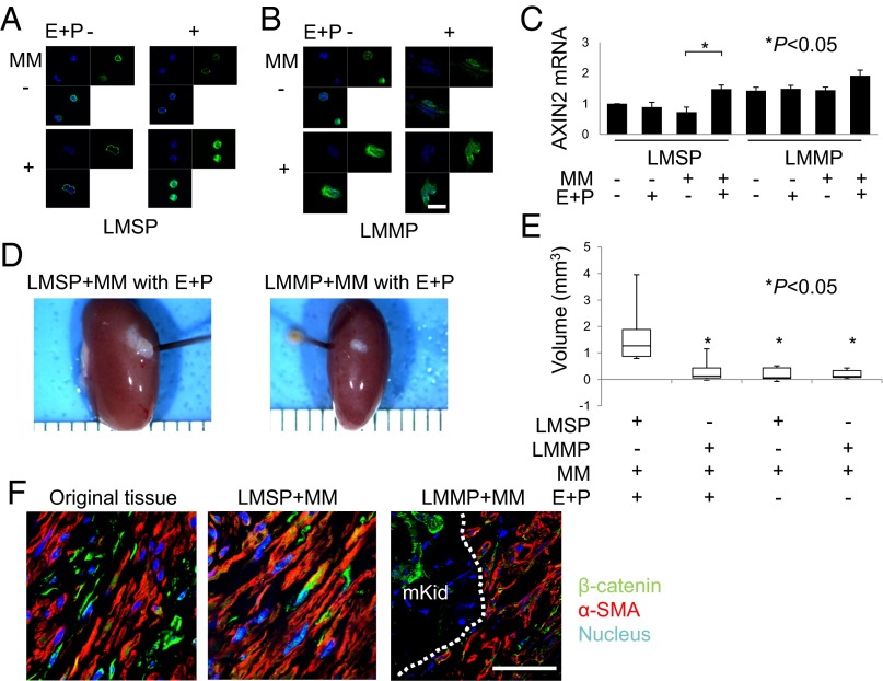
Fig. 2.
E+P indirectly increases WNT signaling activity in LMSP cells. (A and B) Subcellular localization of β-catenin in LMSP or LMMP cells cultured in Transwells with or without MM coculture and in the presence or absence of E+P treatment. Indirect immunofluorescence was performed on LMSP and LMMP cells using anti–β-catenin antibody (1:100; green), followed by Hoechst nuclear counterstaining (blue). The overlay of β-catenin (green) and Hoechst (blue) is shown also. (Scale bar, 20 µm.) (C) Total RNA was extracted from LMSP or LMMP cells cultured in Transwells with or without MM to quantify the expression of AXIN2 by real-time quantitative PCR. Results are expressed as the mean ± SD of AXIN2 relative to GAPDH from four independent experiments. *P < 0.05. (D) Freshly isolated LMSP cells mixed with freshly isolated MM cells are able to generate larger LM tumors than mixes of LMMP-MM cells, with tumor sizes dependent on E+P treatment. Macroscopic visualization of the transplanted site 8 wk after xenotransplantation is shown. (E) Tumor volume of xenografts after 8 wk. Data are shown as mean ± SD. *P < 0.05. (F) Original tissue and generated tumor were analyzed by immunofluorescence. Immunostaining was performed to confirm the presence of β-catenin and α-SMA. Nuclei were stained with DAPI (blue). mKid, mouse kidney. Dotted line indicates the border between mouse kidney and tumor. (Scale bar, 30 µm.)

