Fig. 3.
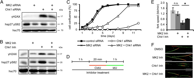
The genotoxic effects caused by depletion or inhibition of Chk1 depend on MK2. (A and B) H2AX phosphorylation upon depletion or inhibition of Chk1 is reduced by codepletion or inhibition of MK2. (A) Cells were depleted of MK2 and Chk1 and harvested 48 h later, and cell lysates were analyzed by immunoblotting. (B) Cells were treated with MK2 inhibitor, Chk1 inhibitor (Chk1 Inh), or DMSO for 12 h, and analyzed by immunoblotting. (C) Cell proliferation after Chk1 depletion is improved by codepletion of MK2. Cells were depleted of MK2 and/or Chk1 and reseeded. At 24 h later, measurement was started (day 1). Cell confluence was measured on subsequent days. Averages of three replicates are shown. (D–F) MK2 inhibition improves the reduced replication fork speed following Chk1 inhibition. (D) Labeling protocol for DNA fiber analysis. Cells were pretreated with MK2 inhibitor, Chk1 inhibitor, or DMSO for 1 h and then pulse-labeled with CldU for 20 min and IdU for 1 h in the continuous presence of inhibitors. CldU and IdU were detected by immunofluorescence in red and green, respectively. (E) Average replication fork speed in dependence of Chk1 and MK2 inhibition. The length of CldU tracks of ongoing forks was used to calculate the replication fork speed (n = 3; *P = 0.0226). (F) Representative images of fibers from cells treated as in D. (Scale bar: 10 µm.)

