Fig. 1.
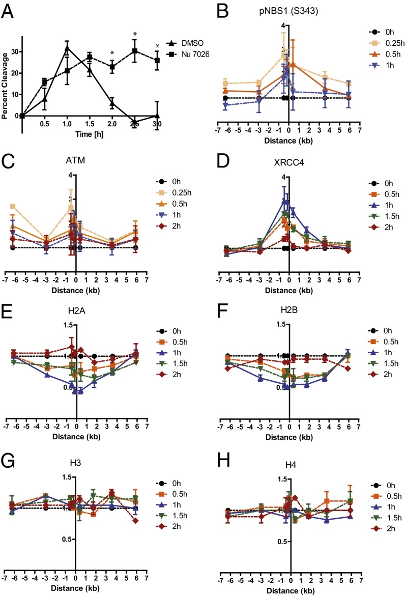
Monitoring repair, DDR protein dynamics at a DSB, and a partial nucleosome disruption in G1 using a tightly regulated ddI-PpoI system. (A) DNA repair measured by quantitative real-time PCR spanning the unique I-PpoI cleave site at chromosome 1 in MCF7 cells stably expressing ddI-PpoI. The DNA-PK inhibitor Nu7026 was added 1 h before 4-OHT and was present during treatment and recovery. Time indicated is hours after the addition of 4-OHT. Data are shown as mean ± SEM of three independent experiments. *P < 0.05. (B–D) ChIP assay showing the recruitment of (B) pNBS1 (S343), (C) ATM, and (D) XRCC4 to the I-PpoI–induced DSB at chromosome 1 in MCF7 cells stably expressing ddI-PpoI. Time indicated is hours after the addition of 4-OHT. The I-PpoI cleavage site on chromosome 1 is located at distance 0. Data are shown as mean ± SEM of two independent experiments. The y-axis displays the fold change in relative occupancy normalized to the control. (E–H) Partial nucleosome disruption occurs in G1-arrested cells. ChIP of (E) histone H2A, (F) histone H2B, (G) histone H3, and (H) histone H4 in MCF7 cells stably expressing ddI-PpoI as described in B. Cells were cultivated in medium containing 0.1% FBS for 24 h before DSB induction.

