Fig. 6.
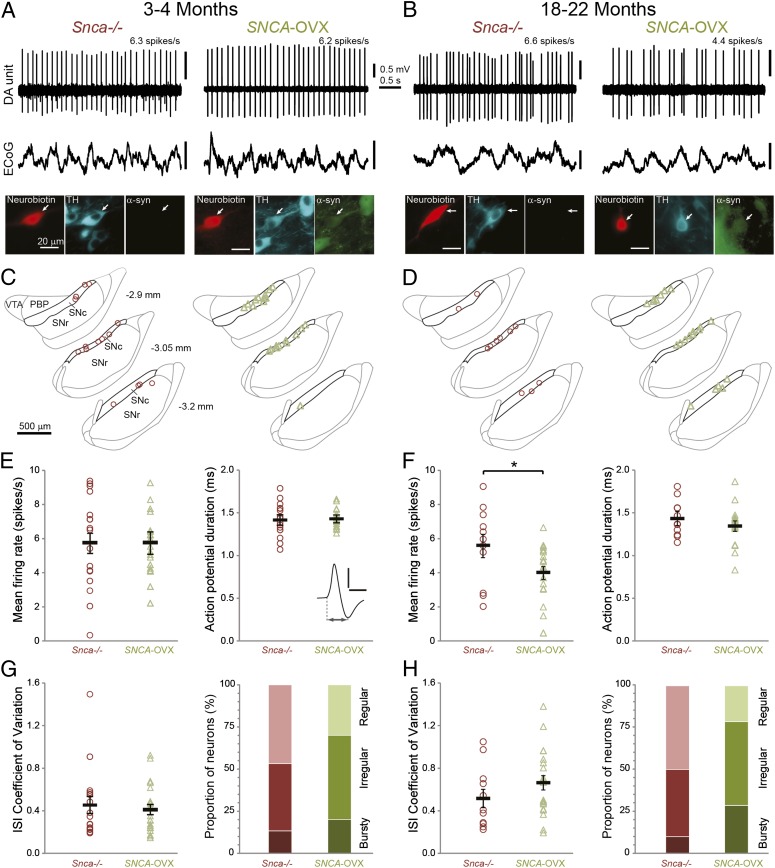
Spontaneous firing of identified dopaminergic neurons in the substantia nigra in vivo. Unit activity of individual dopamine neurons recorded in (A) 3- to 4-mo-old and (B) 18- to 22-mo-old Snca−/− and SNCA-OVX mice during robust slow-wave activity in electrocorticograms (ECoG). Neurons were juxtacellularly labeled with Neurobiotin, verified as dopaminergic by TH expression, and tested for expression of α-syn. (C and D) Locations of identified dopamine neurons in (C) 3- to 4-mo-old and (D) 18- to 22-mo-old Snca−/− (red) and SNCA-OVX (green) mice (dorsal top, lateral right) grouped at three rostro-caudal levels (distances from bregma shown left). PBP, parabrachial pigmented nucleus of the VTA; SNc, substantia nigra pars compacta; SNr, substantia nigra pars reticulata; VTA, ventral tegmental area. [Adapted from ref. 23.] (E and F) Mean firing rates and action potential durations of SNc dopamine neurons in (E) 3- to 4-mo-old (n = 17, Snca−/− and 22 neurons, SNCA-OVX) and (F) 18- to 22-mo-old mice (n = 11 and 21; *P < 0.05). (Inset) Average wideband-filtered action potential from an SNCA-OVX mouse indicating duration measurement (scale 0.5 mV, 1 ms). (G and H) Mean interspike interval (ISI) coefficients of variation and the proportions of SNc neurons exhibiting each firing mode in (G) 3- to 4-mo-old mice (n = 15 and 20 neurons analyzed for mode in Snca−/− and SNCA-OVX) and (H) 18- to 22-mo-old mice (n = 10 and 14 neurons). The firing pattern did not differ significantly with genotype. In E–H, group means ± SEM are shown in black.

