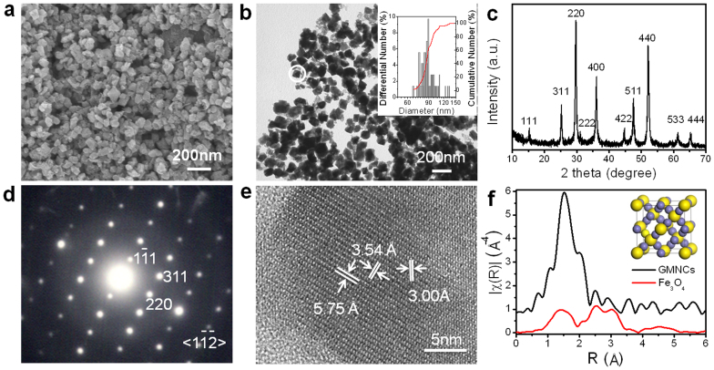
Figure 2. Morphology and phase characterization of the magnetosome-like GMNCs.
(a), FESEM and (b), TEM images show the morphologies and diameters of the as-prepared GMNCs synthesized in EG at 160°C for 2 h. Inset, the size histogram. (c), XRD pattern demonstrated the pure greigite phase. (d), SAED and (e), HRTEM are taken from the selected nanocrystal in (b), exhibiting high crystalline. (f), Fourier transformed XAFS functions in the R domain further illuminated the fcc Fe3S4 phase. Inset shows the cell structure of fcc Fe3S4.

