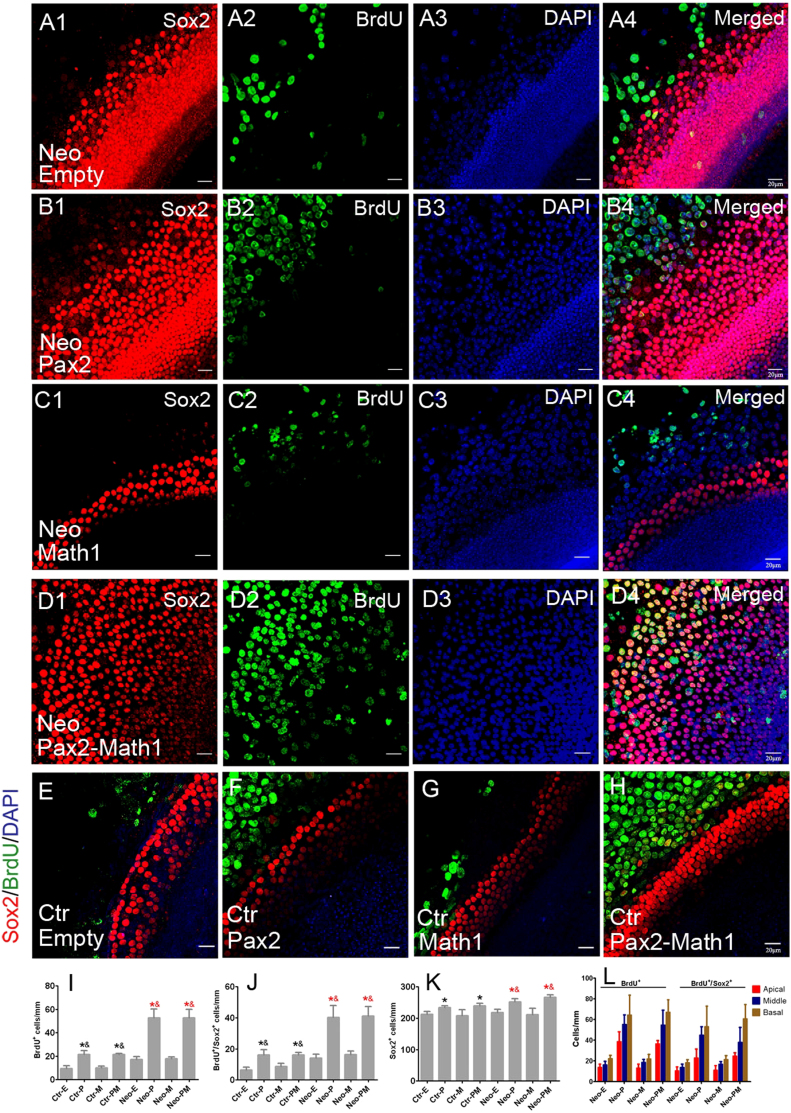
Figure 2. Forced Pax2 overexpression stimulated proliferation of supporting cells.
(A–D) Double immunofluorescence of BrdU and Sox2 in neomycin-damaged cochlear epithelium from the Ad-Empty, Ad-Pax2, Ad-Math1, and Ad-Pax2-IRES-Math1 groups. (E–H) Double immunofluorescence of BrdU and Sox2 in non-damaged cochlear explants from the Ad-Empty, Ad-Pax2, Ad-Math1, and Ad-Pax2-IRES-Math1 groups. Statistical data (I–K) showed that Pax2 overexpression increased the number of BrdU+, Sox2+, and BrdU+/Sox2+ cells. * (red): p < 0.05 vs. Ctr-E; & (red): p < 0.05 vs. Ctr-M; *(black): p < 0.05 vs. Neo-E; & (black): p < 0.05 vs. Neo-M. (L) The distribution of BrdU+ and BrdU+/Sox2+ cells in different regions throughout the damaged cochlea. Scale bars: 20 μm.

