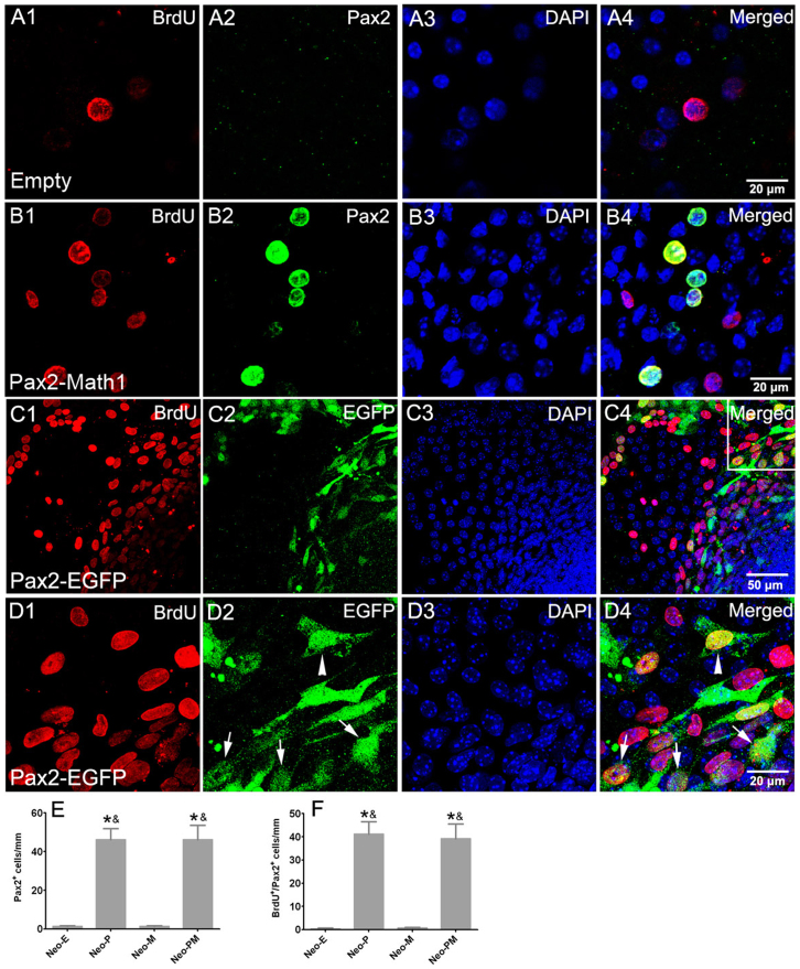
Figure 3. Forced expression of Pax2 stimulated cell proliferation.
(A and B) Double immunofluorescence of Pax2 and BrdU showed that most of Pax2 positive cells were proliferating in the neomycin-damaged cochlear epithelium in Ad-Pax2-IRES-Math1 group. Statistical data (E–F) showed that the number of Pax2+ and BrdU+/Pax2+ cells increased in Ad-Pax2 and Ad-Pax2-IRES-Math1 groups. *: p < 0.05 vs. Neo-E; &: p < 0.05 vs. Neo-M. (C) Double labeling of EGFP and BrdU in damaged cochlear epithelium at 7 days after Ad-Pax2-IRES-EGFP infection. (D) Higher magnification image of (C). Arrows pointed to proliferating supporting cells beneath pre-existing hair cells. The arrowhead pointed to a proliferating fibroblast cell. Scale bars: 20 μm in (A, B, and D); 50 μm in (C).

