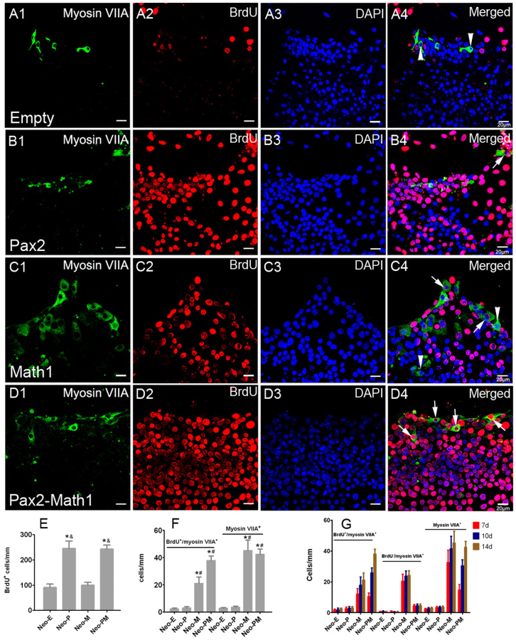
Figure 4. Forced Math1 expression promoted hair cell formation in the lesser epithelial ridge (LER).
(A–D) Double immunofluorescence of BrdU and myosin VIIA in the LER of neomycin-damaged cochlear epithelium at 2 weeks after adenovirus incubation. Myosin VIIA was used as a marker of hair cells. The arrows pointed to BrdU+/myosin VIIA+ hair cells that means hair cells formed by mitotic regeneration. The arrowheads pointed to BrdU−/myosin VIIA+ cells that mean hair cells formed by direct transdifferentiation in the LER. Statistical data (E–F) showed that Math1 expression promoted hair cell formation in the LER in both Ad-Math1 and Ad-Pax2-IRES-Math1 groups. *: p < 0.05 vs. Neo-E; #: p < 0.05 vs. Neo-P; &: p < 0.05 vs. Neo-M. Data in (G) showed the number of BrdU+/myosin VIIA+, BrdU−/myosin VIIA+, and total myosin VIIA+ cells in the LER at different times after adenovirus incubation. Scale bars: 20 μm.

