
Figure 4.
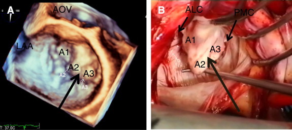
3D TEE- surgery correlation of a patient with severe prolapse of A2 and A3 (two large arrows) in a degenerative (myxomatous) mitral valve. (A) 3D TEE surgical view of the mitral valve with severe prolapse of the anterior mitral leaflet at A2, A3 segments. (B) Corresponding view of surgical demonstration of mitral valve showing severe prolapse of the A2 and A3 due to elongated chorda. AOV = aortic valve, LAA = left atrial appendage, A1 = lateral segment of the anterior mitral leaflet, A2 = middle segment of the anterior mitral leaflet, A3 = medial segment of the anterior mitral leaflet, ALC = anterolateral mitral valve commissure, PMC = posteromedial mitral valve commissure.