
Figure 12.
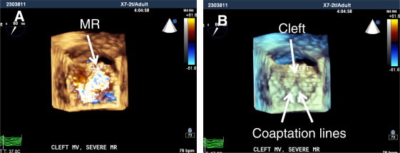
3D TEE full-volume acquisition of the mitral valve (MV) in same patient of Fig. 10, showing surgical view of the cleft MV. (A) mitral valve in systole demonstrating severe mitral regurgitation (MR) mostly origination from the cleft site of anterior mitral leaflet (arrow). Note: in older patients with cleft, due to secondary changes in coaptation line with posterior mitral leaflet (PMVL) and dilatation of the mitral annulus, degree of MR will progress. (B) Same view of mitral valve with color suppress to visualize site of the cleft and line of coaptation with PMVL (arrows).