
Figure 13.
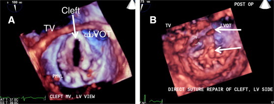
Pre and post- op comparison of the left ventricular (LV) side of the mitral valve MV) in same patient with cleft MV. (A) Pre-op, 3D TEE zoom-mode acquisition of the MV at systole showing cleft of the anterior mitral leaflet oriented towards LVOT. (B) Post-op, same view of the MV in systole after direct suture closure of the cleft (arrows).