
Figure 15.
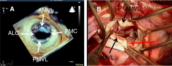
3D TEE-surgery correlation of the same patient of Fig. 14. (A) 3D TEE surgical view of mitral valve (MV) showing diastolic doming and mildly restricted leaflets. (B) Surgical demonstration of the MV prior to repair. Note: calcified free margins of the mitral leaflets. Verrucous non-bacterial endocarditis and erosion of the free margins of mitral leaflets are pathognomonics of rheumatic involvement of the MV (red arrow). 3D TEE is not very sensitive to detect calcification of the leaflets due to poor spatial resolution. ALC = anterolateral commissure, PMC = posteromedial commissure, AMVL = anterior mitral valve leaflet, PMVL = posterior mitral valve leaflet.