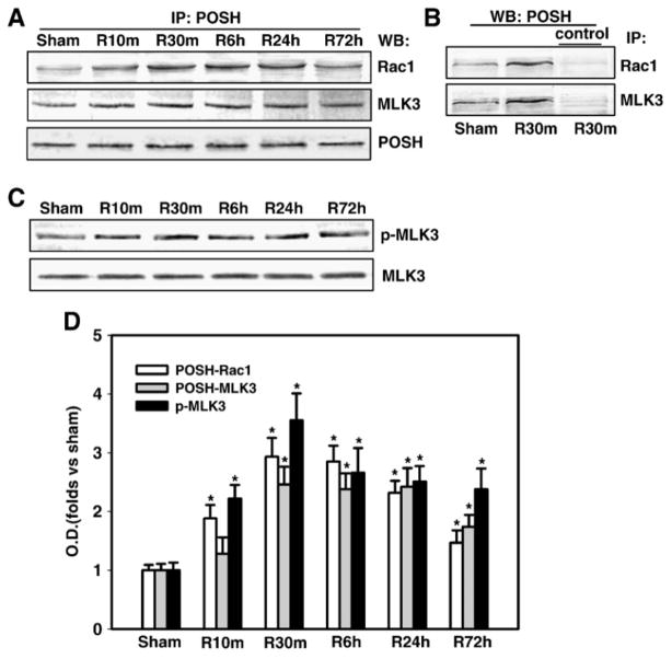
Fig. 2. Time courses of the associations of POSH with Rac1 and MLK3 and phosphorylation of MLK3 in the hippocampal CA1 region after cerebral ischemia.
(A) Homogenates from the CA1 region at various time points after reperfusion (sham, 10 min, 30 min, 6 h, 1 and 3 days) were immunoprecipitated (IP) with anti-POSH antibody, then separately blotted (WB) with anti-Rac1, MLK3 or POSH antibody. (B) In reciprocal co-immunoprecipitation experiments, homogenates were subjected to immunoprecipitation with anti-Rac1, MLK3 or non-specific IgG (control) and the immunocomplexes were probed for the presence of POSH. (C) Homogenates from the CA1 region at various time points of reperfusion were western blotted with antibody against MLK3 or p-MLK3. (D) Corresponding bands from A&C were scanned and the optical density (OD) was represented as folds versus sham control. Data are expressed as means ± SD from independent animals (n = 4–5), *p < 0.05 versus sham control.

