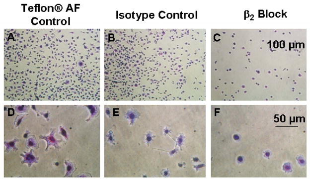
Figure 9.

Phase contrast photomicrographs of results for control and β2 blocks performed on IC-21 cells grown on Teflon® AF surfaces preconditioned with 10% FBS. Cells were incubated without antibodies or with either isotype control or blocking antibodies at a concentration of 100 μg/ml for 30 minutes prior to seeding at a density of 500 cells/mm2. Cells were fixed and stained after one hour. Scale bars are relevant for each row. Images are representative of multiple fields (≥ 3) and multiple replicates (≥ 2) for each test condition.
