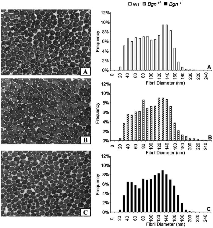
Figure 6.
Representative TEM images of fibril cross-sections and histograms of collagen fibril diameters in (A) WT, (B) Bgn+/−, and (C) Bgn−/− tendons. In all genotypes, most fibrils are well-formed and circular in shape. Some fibrils were slightly abnormally shaped around the perimeter but differences in the frequency of these fibrils were not noted across genotypes. Scale bar 200 nm. Qualitatively in the Bgn+/− a small increase in larger fibrils (longer tail) can be seen. In the Bgn−/−, the density of large fibrils appears to be increased compared to WT.

