Figure 2.

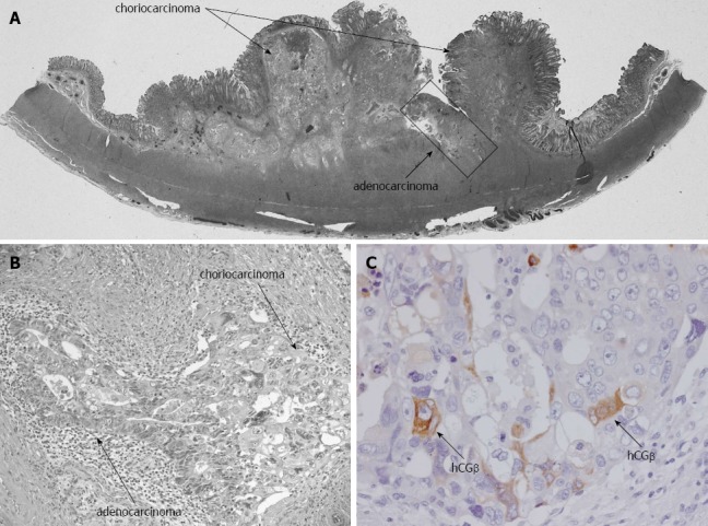
Microscopic findings. A, B: HE staining shows the co-existence of choriocarcinoma and adenocarcinoma cells (A: loupe, B: × 200); C: The tumor cells were positive for β-human chorionic gonadotropin (hCGβ) (× 400).

Microscopic findings. A, B: HE staining shows the co-existence of choriocarcinoma and adenocarcinoma cells (A: loupe, B: × 200); C: The tumor cells were positive for β-human chorionic gonadotropin (hCGβ) (× 400).