Figure 3.
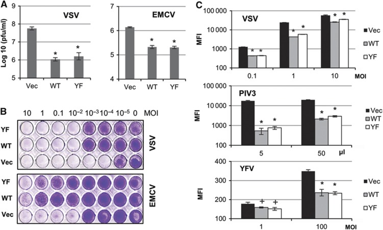
High levels of STAT1, STAT2, and IRF9 proteins protect cells from various RNA viruses in an IFN-independent manner. hTERT-HME1 cells transfected with empty vector (Vec), wild-type STAT1/STAT2/IRF9 (WT) or Y701F-STAT1/STAT2/IRF9 (YF) were used. (A) Cells were infected with 0.1 MOI of VSV or 1 MOI of EMCV, and the infected cells and cell-culture media were collected after 10 h (VSV) or 6 h (EMCV). The infectious viral titres in the collected samples were analysed by plaque assays on Vero cells. The data are represented as means of triplicate infections±s.d. An asterisk (*) represents P<0.01, by two-tailed t-test, compared to cells transfected with empty vector (Vec). (B) Cells were infected with VSV or EMCV (10–10−5 MOI). After 48 h, the surviving cells were fixed with methanol (VSV) or 4% paraformaldehyde (EMCV) and stained with crystal violet. (C) Cells were infected with recombinant viral constructs (VSV, PIV3, or YFV) expressing GFP. After 8 h (VSV) or 48 h (PIV3 or YFV), GFP fluorescence was monitored by FACS analyses. The data are represented as mean GFP intensities (MFI)±s.d. of triplicate infections. An asterisk (*) represents P<0.01 and a cross (+) represents P<0.05, by two-tailed t-test, compared to Vec cells.

