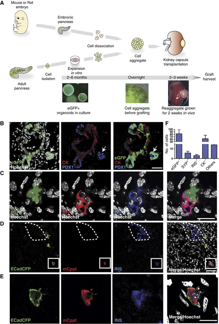
Figure 6.
In vitro expanded organoids from single epithelial cells give rise to endocrine and duct cells when grafted in vivo in a developing pancreas. (A–F) Pancreas organoid cultures were derived from CAGeGFP+ mice or ECadCFP+ mice as described in Figure 3 and Supplementary Figure S3. The cultures were clonally expanded in vitro for 4–6 passages before dissociation into single cells. Dissociated eGFP+ or ECadCFP+ cells were re-aggregated with WT embryonic E13 mouse (B, C) or E14 rat (D, E) pancreas. The re-aggregates were kept on a filter membrane O/N and then grafted under the kidney capsule of nude mice. The grafts were harvested and analysed 2–3 weeks after. The re-aggregates consistently grew and gave rise to pancreatic tissue, as illustrated in Supplementary Figure S7A. (A) Schematic representation of the pancreatic morphogenetic assay. (B) Representative confocal microscopy image showing incorporation of eGFP+ cells (green) into pancytokeratin+ (CK, red) pancreatic duct structures; these eGFP+ cells also express low levels of PDX1 (blue). Other eGFP+ cells (white arrow) aggregated in islet-like structures near the ducts, downregulated CK and expressed high levels of PDX1 (blue). Scale bar: 35 μm. (C) Confocal microscopy demonstrates that cultured eGFP+ (green) cells differentiate into beta cells and express both synaptophysin (SYP, red) and insulin (INS, blue). Scale bar: 20 μm. (D) Confocal microscopy image illustrating mouse Insulin+ Cpeptide+ (INS+Cppt+) cells derived from ECadCFP+-grafted cells. Note that the INS+Cppt+ cells are incorporated into an embryonic rat pancreas, where rat INS+ cells are negative for mouse-specific Cppt staining (dotted line). Scale bar: 30 μm. (E) High magnification of an ECad+INS+Cppt+-grafted cell (D) that displays CFP membrane localization and cytoplasmic staining for INS and mouse Cppt. Scale bar: 20 μm. (F) Histogram showing the average quantification of differentiation of eGFP+ cells engrafted into each re-aggregate under the kidney capsule. Endocrine (SYP+), Insulin (INS+), duct cells (CK+); others: cells expressing neither duct nor endocrine markers. Average number/graft ±s.e.m. n=11 grafts from six independent cultures.

