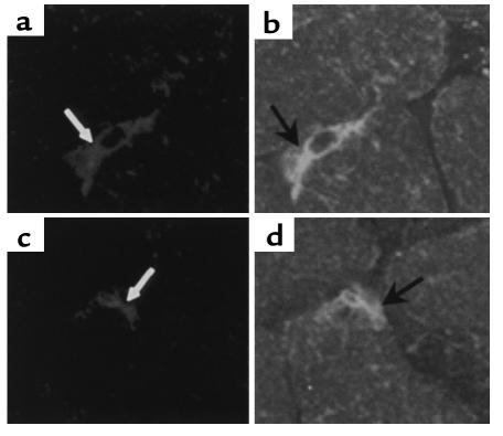
Figure 3.
CD34+ cells are present in ischemic limb vasculature. Confocal images of 10-μm sections of muscle from an ischemic limb of two diabetic mice treated with CM-DiI–labeled CD34+ cells. Sections from muscle collected 18 days after surgery were immunolabeled with FITC-conjugated anti–Tie-2 Ab. (a and b) The same section. (c and d) The same section. Confocal images of CM-DiI–labeled cells (white arrows) (a, c) that have formed vessels and are Tie-2 immunolabeled (black arrows) (b, d).

