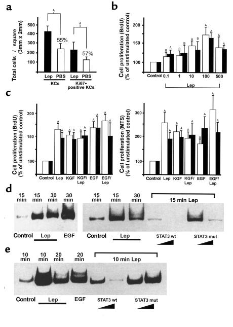
Figure 5.
Effect of leptin on proliferation and STAT3 activation in HaCaT keratinocytes and human primary keratinocytes. (a) Low-magnification photographs (100×) of frozen serial sections were analyzed for total keratinocyte cell numbers or proliferating keratinocytes within the hyperproliferative epithelia from leptin-treated (intraperitoneally) ob/ob mice and PBS-treated (intraperitoneally) ob/ob mice (n = 3), as indicated. This was done by counting hematoxylin-stained keratinocyte nuclei or Ki67-immunostained keratinocyte nuclei in a defined area (3 × 2 mm). Data are expressed as total number of keratinocyte nuclei ± SD (n = 3). AP < 0.01 as indicated by the brackets, percentage of means compared with leptin-treated animals. (b) Dose-dependent effects of human recombinant leptin (0.1–500 ng/mL) on HaCaT (open bars) or primary (filled bars) keratinocyte proliferation. Proliferation was determined by BrdU incorporation. Data are expressed as percentage of unstimulated control. Mean percentage of change in proliferation ± SD are shown (values represent the mean of five assays with a readout of nine wells for each concentration; n = 45). AP < 0.01 compared with control; BP < 0.05 compared with control. (c) Effects of leptin (100 ng/mL), KGF (10 ng/mL), or EGF (10 ng/mL) on HaCaT (open bars) and primary (filled bars) keratinocyte proliferation. KGF and leptin or EGF and leptin were also given simultaneously, as indicated. Proliferation was determined by BrdU incorporation (left panel) or using the MTS reagent (right panel) as described in Methods. Data are expressed as percentage of unstimulated control. Mean percentage of change in proliferation ± SD are shown (values represent the mean of six assays with a readout of nine wells for each condition; n = 54). AP < 0.01 compared with control. (d and e) EMSA. HaCaT keratinocytes (d) and primary keratinocytes (e) were grown to confluence and rendered quiescent by a 24-hour incubation in serum-free medium. Cells were subsequently stimulated with leptin (100 ng/mL) or EGF (10 ng/mL) for the indicated time periods. Nuclear extracts of stimulated cells were isolated as described in Methods. Specificity of binding was confirmed by competition experiments using a 10–100-fold excess of unlabeled wild-type (STAT3 wt) or mutated STAT3 (STAT3 mut) consensus oligo as indicated. Lep, leptin; KC, keratinocyte.

