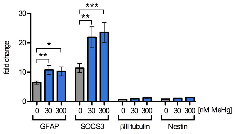
Fig. 3.
MeHg enhances STAT3 target gene expression. Levels of mRNA transcripts were assessed by qRT2PCR in NPCs pretreated for 5 h with 0, 30, or 300 nM MeHg followed by stimulation with CNTF (20 ng/mL) or vehicle for 12 h. Expression levels were normalized to beta-actin expression and standardized to expression levels observed in vehicle treated samples that were not exposed to CNTF. In vehicle treated cultures, CNTF alone caused a 6-fold and 11-fold increase in GFAP and SOCS3 mRNAs, respectively. Exposure to 30 and 300 nM MeHg in the presence of CNTF led to a further increase in GFAP and SOCS3 mRNA expression levels to approximately 10- and 22-fold, respectively. The expression levels of βIII-tubulin, a marker for immature neurons, and Nestin, a marker for uncommitted progenitors, were not significantly altered by CNTF or MeHg treatment. Results are mean fold-change from untreated control +/- SEM from at least 3 independent replicates per condition. * = p < 0.05, ** = p < 0.01 significantly different from positive control by ANOVA and post hoc Bonferroni's test. No changes in any of the genes were observed when NPC were treated with MeHg alone (not shown).

