(COLOR FOR WEB) Fig. 4.
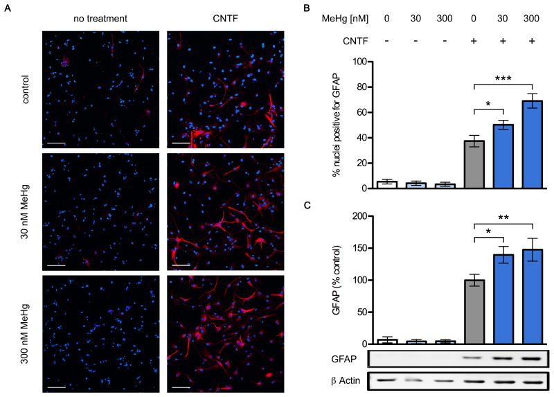
Chronic exposure to low level MeHg enhances glial differentiation. NPCs were grown for two days with or without 20 ng/mL CNTF, in the presence of 30 or 300 nM MeHg or vehicle. Cultures were fixed and GFAP expressing cells were identified using immunofluorescence. Representative images (A), GFAP: red, Hoechst 34580: blue. CNTF increased the proportion of GFAP+ cells, and this was enhanced in the 30nM and 300nM conditions. Also note the more elaborate branching of stained cellular processes in the presence of MeHg (scale bar = 100 μm). Percentage of nuclei expressing GFAP (B), 6 independent experiments, mean +/- SEM. * = p < 0.05, ** = p < 0.01, *** = p < 0.001 significantly different from positive control by ANOVA w/Bonferroni's test. GFAP and β actin protein levels in whole cell lysates were determined using western blots (C). GFAP was quantified by densitometry, normalized to β actin levels, and expressed as percent change from positive control. MeHg significantly increased GFAP protein content in the CNTF-treated conditions, reaching 139 and 148% at doses of 30nM and 300nM, respectively. No effects on GFAP levels were observed in cells not treated with CNTF, regardless of MeHg treatment. Data are mean +/- SEM from 6 independent experiments. * = p < 0.05, ** = p < 0.01 significantly different from positive control by ANOVA w/Bonferroni's test.

