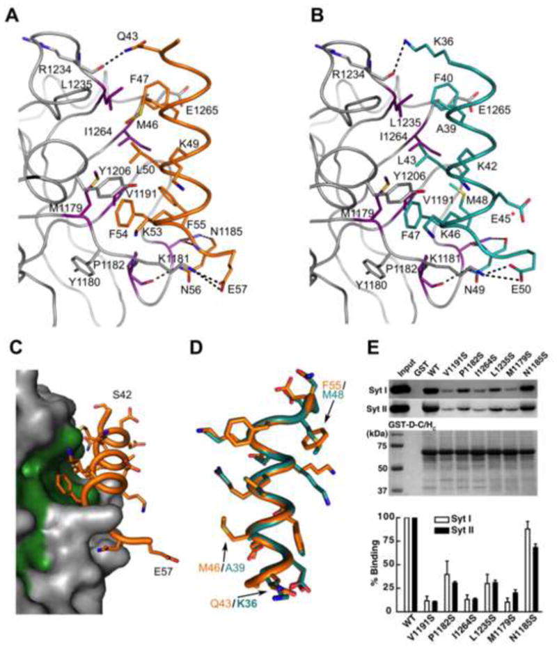
Figure 2. Interactions between BoNT/DC and Syt-II.

(a) A close-up view of the binding interface between BoNT/DC (gray) and Syt-II (orange). All directly interacting residues are shown as sticks. Violet residues at the Syt binding site were selected for mutagenesis studies as described in the following panels. (b) Close-up view of the binding interface between BoNT/DC and Syt-I (teal), with the same coloring as in a. E45, marked with a red star, is the only residue that is different between human and rat Syt-I. E45 is, as seen here, not involved in the binding to the toxin. (c) Surface representation of the Syt-II binding site of BoNT/DC (gray), with bound Syt II (orange) and the hydrophobic residues of BoNT/DC that form the Syt-II binding site shown in green. (d) Comparison of Syt-I and Syt-II bound to BoNT/DC, as seen when superimposing only the BoNT/DC protein chains upon each other. The hydrophobic face of the amphipathic Syt helices, as seen from the BoNT/DC binding site, with the three differences in the binding region between Syt-I and Syt-II highlighted. The other differences between the two isoforms are located in the hydrophilic face of the helix that facing away from the binding site, and are not involved in the binding to BoNT/DC (e) Upper panel: Mutated BoNT/DC-HC were used to pull down native Syt-I/II from rat brain detergent (Triton X-100) extracts. Wild type (WT) BoNT/DC-HC and GST protein were assayed in parallel as controls. Bound Syt-I/II were detected via immunoblot using their specific antibodies. GST-tagged BoNT/DC-HCs used in pull-down assays were visualized by Coomassie blue staining to indicate equal loading. Lower panel: binding of Syt-I/II to BoNT/DC-HC mutants was quantified and normalized to the amount of binding to WT BoNT/DC-HC. See also Figure S1 for a sequence alignment of Syt isoforms. See also Figure S1 for a) CD spectra illustrating that the mutations in BoNT/DC-HC does not induce any major structural changes and b) for a schematic overview of the two binding sites.
