
Figure 5.
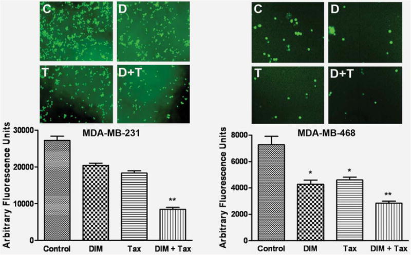
DIM decreased breast cancer cell invasion. Invasion assay showing that DIM-treated breast cancer cells resulted in low penetration through the Matrigel-coated membrane, compared with control cells. MDA-MB-231 and MDA-MB-468 cell were with 25 and 40 μM of DIM, respectively. The bar graphs show the fluorescence values from the invasive cells. The value indicated the comparative amount of invaded cells. C, Control; D, DIM 25 or 40 μM; T, Taxotere (1.0 nM); D+T, DIM + Taxotere; Tax, Taxotere; DIM+Tax, DIM + Taxotere. The experiments were repeated thrice. *, p < 0.05; **, p < 0.01 relatively control. [Color figure can be viewed in the online issue, which is available at wileyonlinelibrary.com.]