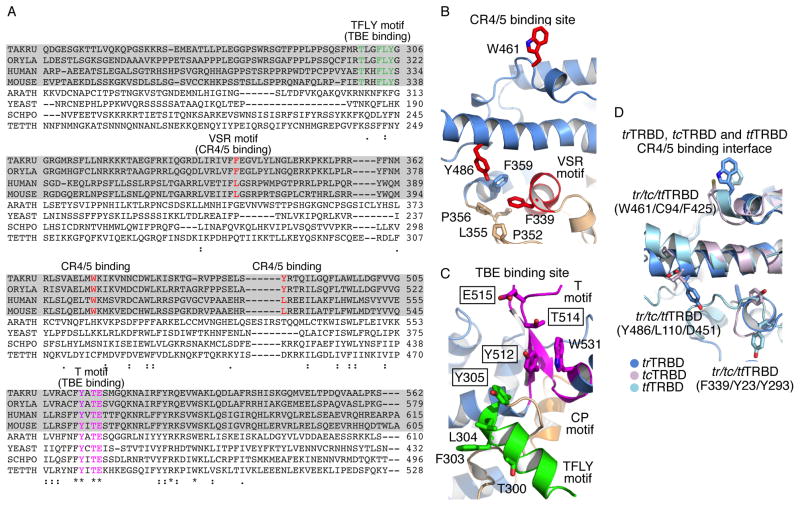
Figure 3. TRBD and N-terminal linker sequence and structural conservation.
A) Sequence alignment of the N-terminal linker and TRBD with the conserved TFLY (green), VSR (red) and T (magenta) motifs shown in color. B and C) Portions of the trTRBD structure highlighting B) the VSR (red) and adjacent residues involved in CR4/5 binding and C) the T (magenta) and TFLY (green) motifs involved in TBE binding. Conserved residues mutated in this study are shown in boxes. D) Structural overlay of trTRBD (blue), ttTRBD (cyan) and tcTRBD (violet) CR4/5 binding surface showing structural and amino acid conservation.

