
Figure 6.
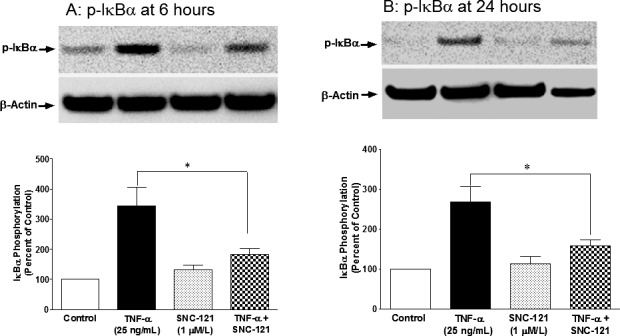
TNF-α–induced phosphorylation of IκBα in ONH astrocytes at 6 (A) and 24 (B) hours. ONH astrocytes were starved in serum-free medium overnight. Cells were then pretreated with SNC-121 (1 μmol/L) for 15 minutes followed by TNF-α (25 ng/mL) treatment for 6 or 24 hours. Cell lysate (15-μg protein) was analyzed by Western blotting using selective anti–phospho-IκBα antibodies followed by incubation with appropriate secondary antibodies (HRP-conjugated; dilution 1:3000). The signal was captured using enhanced chemiluminescent reagent and a commercial imaging system (Bio-Rad). Data are expressed as mean ± SE. *P < 0.05. n = 5 to 6.