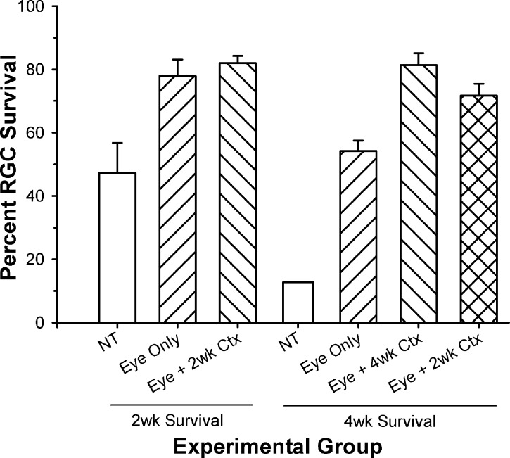
Figure 4.
Comparison of the percent ganglion cell survival in the AC for animals receiving different treatment strategies, and either 2- or 4-week survival periods. At 2 weeks after injury/treatment, there was a significant increase in ganglion cell survival following treatment of the eye alone and the eye + cortex (P < 0.05) compared to NT. Following a 4-week survival period, all treatment conditions enhanced ganglion cell survival significantly relative to NT (P < 0.05), with the DT animals showing a significant increase over those receiving treatment of the eye alone (P < 0.05). Although those receiving only 2 weeks of treatment, followed by an additional 2-week survival period without treatment, showed a slight reduction in ganglion cell survival in the AC, they were not different from those animals receiving 4 weeks of treatment (P > 0.05). The 2-week survival data are from the study of Weber et al.15

