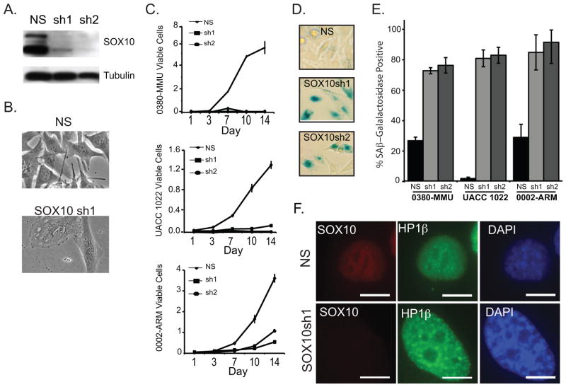
Figure 1.
SOX10 depletion triggers arrested growth and melanoma cell senescence
A. Western blot analysis of SOX10 expression in 0380-MMU cell lysates stably transduced with a non-silencing control hairpin (NS) or SOX10-specific lentiviral RNAi hairpins (sh1 and sh2). B. Phase-contrast images of 0380-MMU stable cell lines, 1 week post-infection with NS or SOX10sh1. C. Proliferation assays for 0380-MMU, UACC 1022, and 0002-ARM cell lines transduced with NS, sh1, or sh2. Cells were counted in triplicate 1 to 14 days after plating, and proliferation assays were performed at least twice with subsequent infections. D. Representative images of 0380-MMU cells stained for SA-β-Gal activity one week post-infection with NS, SOX10sh1 and SOX10sh2. E. Significant increases in the percentage of SA-β-Gal positive cells were seen in SOX10sh lines relative to NS lines, as determined by t-test (for 0380-MMU, sh1 p<7.32×10⁻⁵, sh2 p<7.95×10⁻³; for UACC 1022, sh1 p<5.49×10⁻¹¹, sh2 p<2.17×10⁻⁸; for 0002-ARM, sh1 p<2.90×10⁻⁵, sh2 p<8.80×10⁻⁵). F. Stably transduced UACC 1022 cell lines stained with anti-SOX10, anti-HP1β antibodies and DAPI nuclei counterstain. HP1β staining indicated the presence of senescence-associated heterochromatin foci in SOX10sh1 cells. Scale bar = 10 μm.

