
Figure 10.
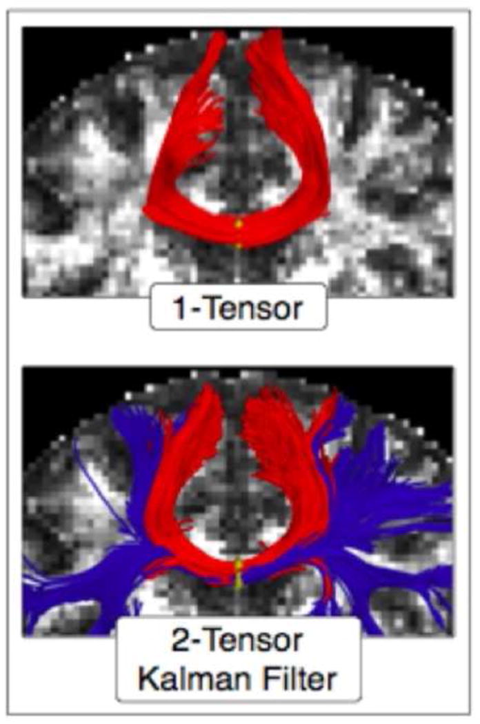
Part of the corpus callosum fibers, where seeding was done in the mid-sagittal plane of the corpus callosum. Top figure shows the tracing using the standard single-tensor model and bottom figure shows tracts generated with the two-tensor model.