
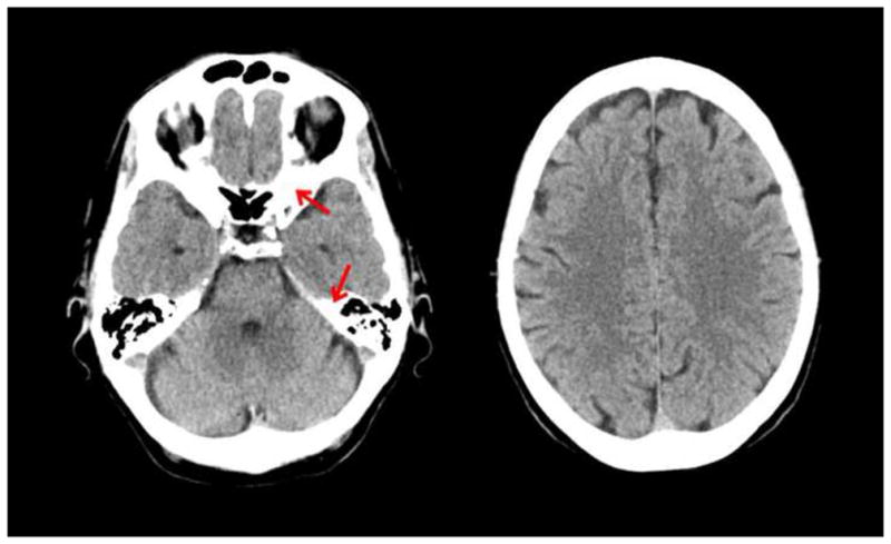
Figure 2.
CT scan of a normal brain. Left side is at the level of the temporal lobe where bone can be seen as white areas (see red arrows). Right side is at the level of the frontal lobe. (Courtesy of Amir Arsalan Zamani, M.D.)
Official websites use .gov
A
.gov website belongs to an official
government organization in the United States.
Secure .gov websites use HTTPS
A lock (
) or https:// means you've safely
connected to the .gov website. Share sensitive
information only on official, secure websites.
CT scan of a normal brain. Left side is at the level of the temporal lobe where bone can be seen as white areas (see red arrows). Right side is at the level of the frontal lobe. (Courtesy of Amir Arsalan Zamani, M.D.)