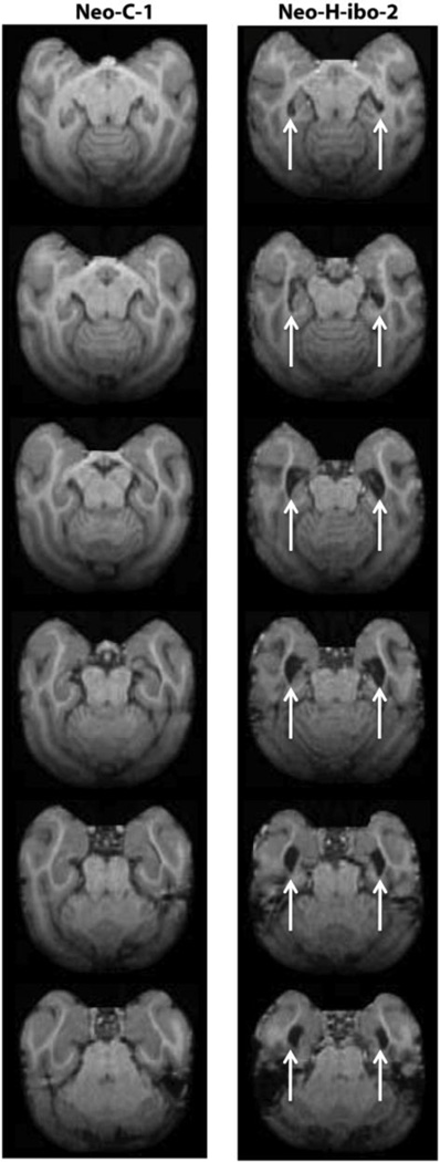
Figure 1.
Transverse T1-weighed images through the brain of a normal control monkey (Neo-C-1) on the left and a monkey with a neonatal lesion of the hippocampus (Neo-H-ibo-2) on the right. Comparisons between the two series of images clearly illustrate the 67% reduction in hippocampal volume in case Neo-Hibo-2, estimated from the one year post-surgical scan (see Table 2). Arrows point to increased lateral ventricle due to volume reduction of the hippocampus.

