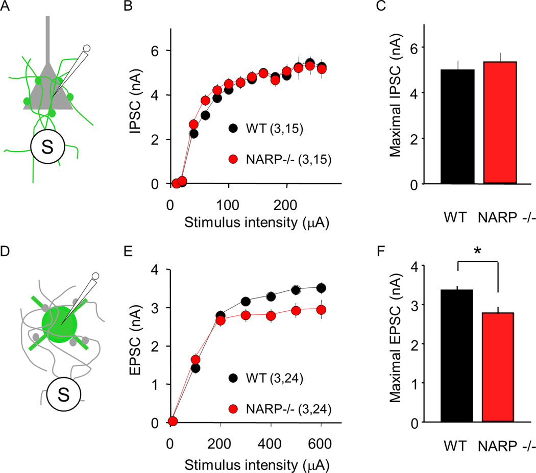
Fig 3. Normal inhibitory input onto pyramidal cell but reduced excitatory input onto FS (PV) INs in NARP−/− mice.
A–C. Extracellularly-evoked IPSCs (eIPSCs) recorded in pyramidal neurons are normal in P35 NARP−/− mice. A. Pharmacologically-isolated eIPSCs were recorded in layer II/III pyramidal neurons, evoked by extracellular stimulation of the underlying layer IV. B. Input-output relationship for eIPSCs in NARP−/− (red) and WT controls (black). C. Maximal IPSC computed by averaging eIPSC amplitudes evoked by the 3 largest stimulus intensities. D–F. Extracellularly- evoked EPSCs (eEPSCs) recorded in FS (PV) INs are reduced in P35 NARP−/− mice. D. Pharmacologically-isolated eEPSCs were recorded in layer II/III FS (PV) INs, evoked by extracellular stimulation of the underlying layer IV. E. Input-output relationship for eEPSCs in NARP−/− (red) and WT controls (black). F. Maximal EPSC computed by averaging eEPSC amplitudes evoked by the 3 largest stimulus intensities. Number of mice and neurons in parentheses in B and E. *=p<0.02; t-test.

